Many people first hear the words “brain aneurysm” in a scan report. The scan was often done for something else: a headache, a dizzy spell, a head injury. Suddenly there is a frightening word on the page, and a long wait before the next appointment.
If this is where you are, this article is for you. It explains what the finding usually means, why some aneurysms are watched rather than treated straight away, how doctors decide when treatment is worth its risks, and what the main treatments involve. It cannot tell you what your own scan means. That depends on details only your treating team can see.
First, what has actually been found?
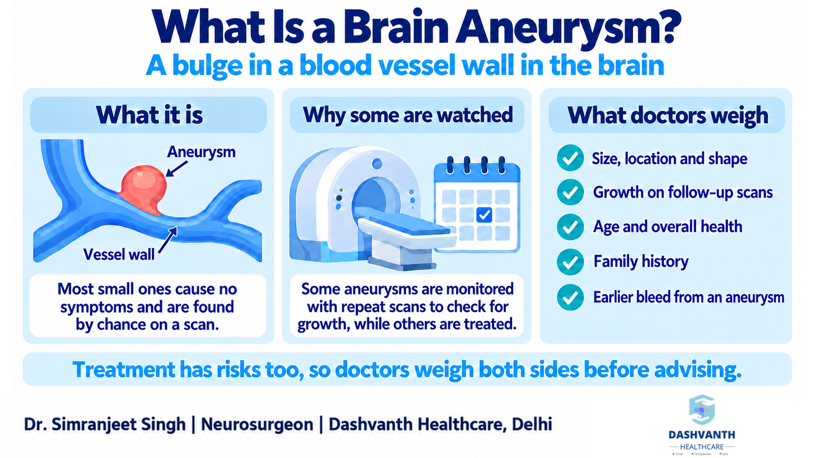
A brain aneurysm is a bulge in the wall of a blood vessel in the brain. Rupture is uncommon, but when it happens it causes bleeding around the brain, called a subarachnoid haemorrhage. NHS: Brain aneurysm
Most small aneurysms cause no symptoms and are found by chance. Whether an aneurysm is watched with regular scans or treated depends on the individual aneurysm and the individual person. AHA/ASA 2015 guideline
Larger aneurysms can sometimes press on nearby nerves. This may cause pain around the eye, double vision or other vision changes, or numbness in the face. These symptoms need medical assessment, but they are not the same as a rupture.
Why “watch and scan” is a real treatment plan
Hearing “we will monitor it” can feel like being told nothing is being done. Monitoring is an active plan. Treatment itself carries risk, so doctors weigh the risk of leaving an aneurysm against the risk of treating it.
The American Heart Association and American Stroke Association guideline says periodic imaging should be considered for unruptured aneurysms managed without treatment, because growth may increase the risk of rupture. It also says that aneurysms shown to enlarge during follow-up should generally be offered treatment, unless other health problems make treatment too risky. AHA/ASA 2015 guideline
CT angiography and MR angiography are the scans commonly used to find and follow aneurysms. AHA/ASA 2015 guideline
What doctors weigh when deciding about treatment
There is no single number that decides this. The guideline lists the factors to weigh together:
- the size, location and shape of the aneurysm
- whether it has been growing
- the person’s age
- any earlier bleed from an aneurysm
- a family history of aneurysms or subarachnoid haemorrhage
- whether there is more than one aneurysm
- other conditions that raise bleeding risk
Doctors also consider the person’s overall health, and what the person wants once the options are explained. Two people with a similar-looking report can reasonably receive different advice. Some teams use risk scoring tools to organise the discussion, but these do not replace it.
Coiling and clipping: how they differ
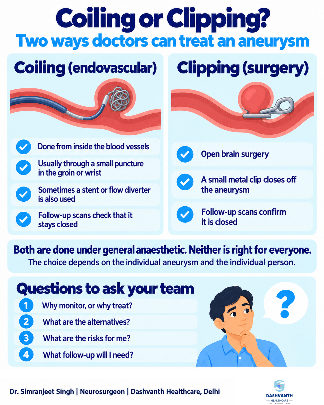
Both are done under general anaesthetic. NHS
Clipping. This is open surgery. A small metal clip is placed across the neck of the aneurysm to stop blood flowing into it.
Endovascular coiling. This is done from inside the blood vessels, usually through a small puncture in an artery in the groin or wrist. A catheter is guided up to the aneurysm, and soft coils are placed inside it to slow blood flow and encourage clotting. For some aneurysms, an additional device is used: a stent placed in the parent vessel to hold the coils in position (stent-assisted coiling), or a flow diverter, a fine mesh tube placed in the parent vessel that redirects blood flow away from the aneurysm. Whether such a device is needed depends on the shape of the aneurysm. NHS
Both are recognised options, and neither is better in every situation. Which one is chosen depends on the individual aneurysm and the individual person: its size, shape and position, the person’s age and general health, and their own wishes once the options have been explained. Some aneurysms are suited to one approach and not the other. This is why the decision is often made by a team that includes both neurosurgeons and neurointerventional specialists. AHA/ASA 2015 guideline
Risks and limitations, stated plainly
Any treatment for an aneurysm can cause complications. They include stroke, bleeding, damage to nearby blood vessels or nerves, and, rarely, death. The chance of a complication depends on the aneurysm and on the person, so a single number would be misleading. Ask your treating team how the risk applies to you.
Endovascular treatment involves X-ray imaging, so radiation exposure should be explained as part of consent. AHA/ASA 2015 guideline
Treatment lowers the risk of rupture but does not remove every risk. An aneurysm can come back after coiling, and a treated aneurysm is not guaranteed to be gone for good.
After a decision: follow-up is part of the treatment
If you are being monitored, you will have repeat scans at intervals your team chooses. Keep those appointments, even when you feel well.
If you have had treatment, imaging afterwards confirms how completely the aneurysm has been closed, because a completely and an incompletely closed aneurysm carry different risks. AHA/ASA 2015 guideline
Driving and work. Ask your doctor about driving, travel and returning to work for your situation. Advice differs from person to person.
What you can do in the meantime
Smoking is linked to the formation of aneurysms, and high blood pressure may contribute to their growth and rupture. The guideline advises quitting smoking and treating high blood pressure. AHA/ASA 2015 guideline The NHS also lists a close relative with an aneurysm and drug use such as cocaine among the risk factors. NHS
Screening is not for everyone. The guideline suggests offering a scan to people with two or more close family members who have had aneurysms or subarachnoid haemorrhage. If this describes your family, ask your doctor whether screening is appropriate. AHA/ASA 2015 guideline
When this is an emergency
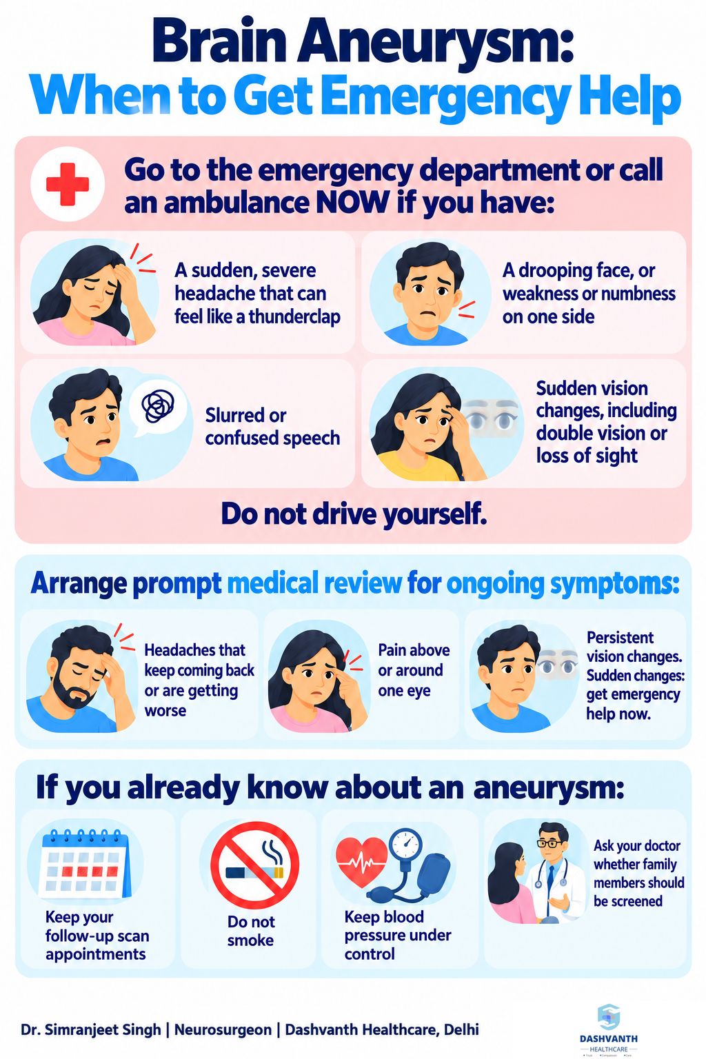
The main warning sign of a rupture is a sudden, severe headache that can come on like a thunderclap. NHS Go to the nearest emergency department or call an ambulance immediately if you have:
- an extremely painful headache that comes on suddenly or will not go away
- a drooping face, or arm or leg weakness or numbness on one side
- slurred or confused speech
- sudden loss of vision or blurred vision
Do not drive yourself. This applies whether or not you already know about an aneurysm. For more on stroke-type warning signs, read Stroke Warning Signs: Why Minutes Count.
Questions worth asking your treating team
- How big is the aneurysm, where is it, and what does its shape look like?
- What are the reasons to watch it, and what would make you recommend treatment?
- How often will I need scans, and which type?
- If treatment is advised, why that option, and what are the alternatives?
- What are the risks for someone of my age and health?
- What happens after treatment, and what follow-up will I need?
- What should I do about blood pressure, smoking and other health conditions?
- Should my family members be screened?
You can read more about neurovascular care on the expertise page.
A word of reassurance
An aneurysm on a scan is a finding to take seriously, not a reason to panic. Some are simply followed with scans, and for those that need treatment there are established options. Understanding the plan, keeping your follow-up appointments and knowing the emergency signs puts you in a much stronger position.
If you have been told you have a brain aneurysm, you may bring your scans and reports to your appointment, along with the questions above.
Dr. Simranjeet Singh
M.Ch. Neurosurgery (AIIMS, New Delhi)
Director, Dashvanth Healthcare, Delhi.
Consultant Neurosurgeon, Max Smart Super Speciality Hospital, Saket.
Learn more about Dr. Simranjeet Singh.
The illustrations in this article are digitally created for representation and education.
This article is for general education and does not replace a personal medical consultation.